· 2 min read
White Matter Hyperintensities Study
New interactive sample study showcasing the results of our AI software on White Matter Hyperintensities (WMH) segmentation task.
New Example on Hetalox Demo Page
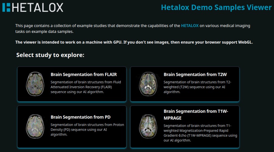
The Hetalox demo page now features a sample study showcasing the results of our AI software on the White Matter Hyperintensities (WMH) segmentation task. You can visit the Hetalox Demo Page to check it out.
White Matter Hyperintensities?
White Matter Hyperintensities (WMH) are brain white matter lesions visible on fluid-attenuated inversion recovery (FLAIR) MRI scans, predominantly in adults and older individuals, with a growing number of lesions throughout the lifespan. They are associated with cognitive decline, dementia, and an increased risk of stroke. Demyelinating lesions, which can appear similar to white matter hyperintensities, typically occur in multiple sclerosis (MS) and are often found in young women, who are predominantly affected by the disease.
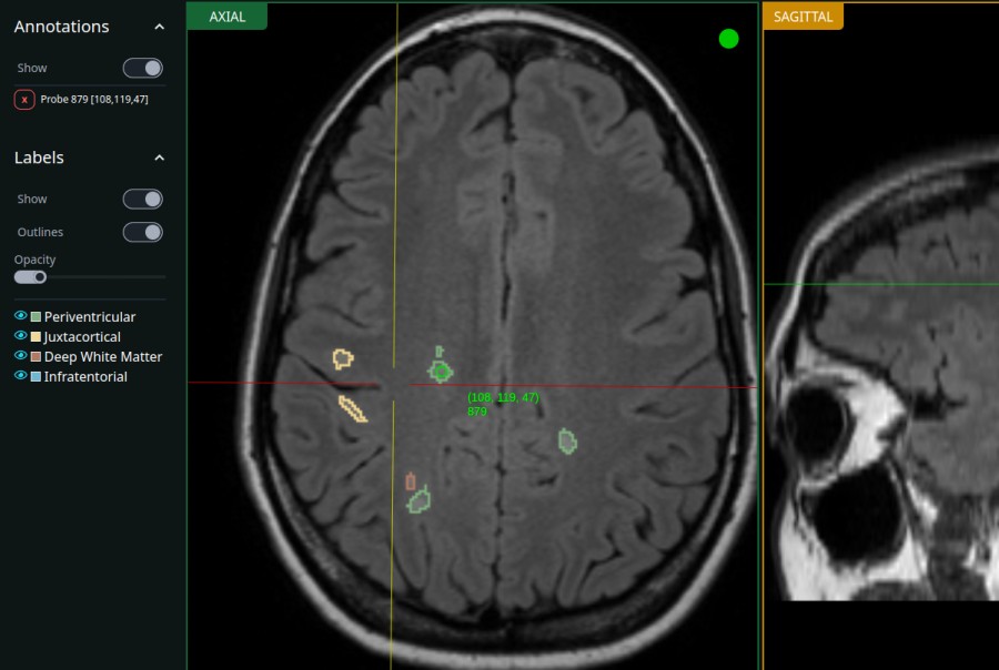
Hetalox can automatically segment WMH on MRI scans, which can help in the early detection and monitoring of these lesions, as well as study these changes over time. Our AI model performs WMH segmentation from 3D and 2D FLAIR sequences, providing repeatable results without the need for time-consuming manual corrections.
Additionally, we color-code WMH localizations to make it easier to visualize and interpret the results. Localization areas include periventricular, juxtacortical, infratentorial, and deep white matter regions. Our platform also provides a quantitative analysis of WMH volume, which can be used to track the progression of the disease and reduce the time and effort required by medical experts.
Additional reads
Here are a few resources to learn more about WMH and their clinical importance: